Japan guide · Tissue engineering
Cell sheet engineering: why cornea worked and heart did not
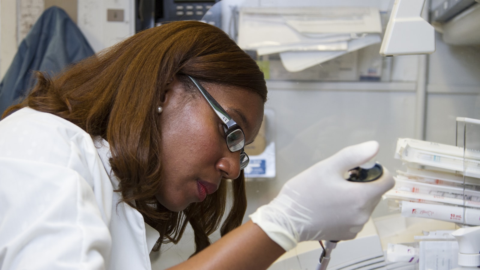
Photo: National Cancer Institute / Unsplash
In September 2015, Terumo's HeartSheet, a cell sheet laid on the surface of the heart, won conditional and time-limited approval in Japan. In July 2024 the review for full approval concluded that efficacy had not been shown, and sales ended. A cell sheet for the cornea, meanwhile, is an approved product today. Why did the same technology end up in two places?
A cell sheet is a way to harvest cultured cells as one intact layer. Instead of detaching cells one by one with enzymes, you lower the temperature of the culture dish and the cells lift off together. How mature the technology is and whether the therapy works are separate questions. Each tissue asks the sheet to do a different job, and measures success differently.
This guide is for researchers, and equally for buyers, sales staff and quality people who handle culture dishes, scaffolds and organoid kits. It compares cell sheets with scaffolds, organoids and bioprinting by strength and weakness, so you know which questions to ask before you choose or recommend one.
The cornea sheet became a product and the heart sheet did not
Same sheet technology, two outcomes. The split comes down to the tissue.
For the cornea, Nishida and colleagues reported in the New England Journal of Medicine in 2004 on sheets of epithelial cells grown from the patient's own oral mucosa and transplanted onto a damaged corneal surface. Vision improved in four patients, and no complications were reported. In March 2020, Nepic, an autologous cultured corneal epithelium from J-TEC (Japan Tissue Engineering, a Fujifilm subsidiary), was approved as the first ophthalmic regenerative medical product (再生医療等製品) in Japan.
For the heart, Terumo's HeartSheet took a sheet of cells made from the patient's own skeletal muscle and applied it to the heart surface. It was approved in September 2015 under conditional and time-limited approval (条件及び期限付承認). The condition was a post-marketing survey covering every patient treated. The time limit was five years, extended by another three in 2019, and Terumo applied for full approval in September 2023. On July 19, 2024, an MHLW subcommittee concluded from the post-marketing results and clinical studies that efficacy had not been shown. No new safety problem had been found. On July 24 the Pharmaceutical Affairs Council also found that approval was not appropriate, Terumo ended sales, and the approval was closed out effective July 25.
The cornea is thin and has no blood vessels. The sheet does a direct job there: it covers a damaged surface again. Heart muscle is thick and beats without rest. The skeletal muscle cells were not expected to turn into heart muscle. Instead, factors released from the sheet were expected to support new blood vessels and heart function, an indirect effect. An indirect effect makes the measurement itself harder, because it is not obvious what "it worked" should look like.
Conditional and time-limited approval exists so that post-marketing data can confirm efficacy. HeartSheet is the case where that check came back without confirming it.
"Does this technology work?" is too coarse a question for any cell sheet, so first state whether the target tissue is thin or thick, whether the sheet replaces tissue or acts on its surroundings, and which endpoint will show an effect.
A temperature-responsive dish works only inside a narrow coating window
The core of the toolkit is PIPAAm (poly-N-isopropylacrylamide), a polymer coated on the dish surface. At 37°C it repels water and cells attach. Around 32°C it switches to water-friendly, and the cells lift off on their own. Okano and colleagues reported the mechanism in 1993. CellSeed commercialized it, and Thermo Fisher Scientific sells it as Nunc UpCell (temperature-responsive culture dishes).
Performance is set by a film only about 15 to 20 nanometers thick. In that range, cells attach firmly at 37°C and detach quickly at 32°C or below. Above 30 nm, cells barely attach even at 37°C. Too thin, and detachment cannot be controlled. Too thick, and attachment never happens. A dish outside the window is plain plastic again.
Detachment speed also depends on what the polymer sits on. One report found that a surface grafted onto a porous membrane released the sheet more than twice as fast as flat polystyrene (Kwon et al., 2000).
If you buy or qualify these dishes, ask the maker how coating thickness is controlled lot by lot and what performance it guarantees.
Sheets, scaffolds, organoids and bioprinting differ in what sets the shape
A cell sheet is one way to build tissue in three dimensions, not the only one. The four routes are best told apart by what determines the shape.
In a cell sheet, the cells themselves make the layer. It suits thin tissue and surface coverage, and thickness has to come from stacking sheets.
In a scaffold, the shape and stiffness of the support set the form. Options include collagen sponges, decellularized tissue, synthetic polymers such as PLGA, and hydrogels. Hard tissue such as cartilage gets a stiff scaffold, and soft tissue such as skin gets a soft gel. The weak point is timing. A degradable scaffold has to disappear at the pace at which cells build their own matrix. If it goes first, the structure collapses. If it lingers, it stays behind as a foreign body (scaffolds and biomaterials). Where large numbers of cells are needed, microcarriers, which are beads that carry cells in stirred culture, also count as a kind of scaffold (microcarrier culture (in Japanese)).
Organoids let stem cells organize themselves. Embed stem cells in a basement membrane matrix, add growth factors, and part of the fine structure of intestine, liver or kidney forms spontaneously. Biology decides the form, not a machine, so the idea of printing precision does not apply. The catch is the matrix. Most of the standard matrices come from mouse sarcoma, vary from lot to lot, and are made for research. The more you care about reproducibility, the more lot control of matrix and growth factors decides whether results repeat (organoid culture kits).
Bioprinting reverses the logic and specifies the shape from outside. Extrusion printing tends to lower viability through shear, the force on cells as liquid is pushed out. Inkjet printing gives high resolution but limits which inks you can use. Whether resolution or survival comes first decides the method (bioinks).
Organoids leave shape to biology and gain reproducibility, bioprinting sets shape from outside and gains freedom, and neither is better, so choose by what you rank first.
Thick tissue meets two walls: blood vessels and testing without destruction
Whatever the method, the thicker the tissue, the harder two problems push back.
The first is vascularization. If oxygen and nutrients cannot reach the interior, the center dies. Even for large bioprinted tissue, the lack of a vascular network is described as the fundamental problem (see this news item on larger bioprinted tissues (in Japanese)). Researchers are stacking sheets to build thick tissue that contains vessel networks, and building vessel structures in at the printing stage.
The second is that few ways exist to check tissue without destroying it. Peeling off one sheet and looking at it is easy. A thick stack, or tissue whose cells have grown into a scaffold, has to be sectioned and stained, and that destroys the product. So quality assurance shifts its weight from testing the final product to process records. These are the cell number and viability at seeding, the differentiation markers at each stage of culture, and the lots of scaffold, matrix and growth factors.
The thicker the tissue you aim for, the more quality assurance rests on process records and the less on final-product testing.
What to check when a tissue-engineering approach reaches your desk
Use these questions on papers, maker materials and contract manufacturer proposals.
- Is the target tissue thin or thick, and does it have few blood vessels?
- What is the sheet or the cell expected to do: replace tissue, or act on its surroundings? Which endpoint measures the effect?
- For a scaffold, does the rate at which it degrades match the rate at which the tissue builds its own matrix?
- For organoids, how are lot-to-lot differences in the matrix and growth factors managed?
- For bioprinting, did the team prioritize resolution or viability, and do they measure viability after printing?
- Can the final product be checked without destroying it? If not, what is recorded during the process?
- In that tissue, how far has the clinical record progressed?
Related reading covers manufacturing mesenchymal stem cells and how organoids are used as in vivo efficacy models in drug discovery (in Japanese).
Sources
- A novel recovery system for cultured cells using plasma-treated polystyrene dishes grafted with poly(N-isopropylacrylamide). Journal of Biomedical Materials Research (Okano et al.), Oct 1993.
- Rapid cell sheet detachment from poly(N-isopropylacrylamide)-grafted porous cell culture membranes. Journal of Biomedical Materials Research (Kwon et al.), Apr 2000.
- Corneal reconstruction with tissue-engineered cell sheets composed of autologous oral mucosal epithelium. New England Journal of Medicine (Nishida et al.), Sep 2004.
- News release on the approval and domestic sale of HeartSheet. Terumo, Sep 2015.
- Conditional and time-limited approval system for regenerative medical products. PMDA, Oct 2026.