Japan guide · Quality control · Mycoplasma
Mycoplasma testing: culture, indicator cell and NAT compared

Cells grow more slowly, yields drop, and experiments stop reproducing, yet the medium is clear and its color has not changed. That is when to suspect mycoplasma. A 2015 paper that searched public data from rodent and primate cells (884 studies, 9,395 samples) found mycoplasma-derived sequences in about 11% of the studies.
Mycoplasma is a bacterium with no cell wall, about 0.2 to 0.3 µm across. It changes shape, and it can slip through the 0.2 µm filters used for sterilizing filtration. "We filter-sterilized, so we are fine" does not hold, so the pharmacopeias set a dedicated test apart from the sterility test: USP <63>, Ph. Eur. 2.6.7 and, in Japan, a general information chapter of the Japanese Pharmacopoeia (JP, マイコプラズマ否定試験). The framework of that test changed with the revision of Ph. Eur. 2.6.7 that came into force on April 1, 2026.
QC runs the test, but QA reviews the choice of method, purchasing picks kits and contract labs, and in cell therapy development the schedule from collection to administration constrains the choice. This guide compares the three methods, shows what changed in 2026 and gives an order for choosing.
Why does a visual check tell you nothing about mycoplasma?
Because there is almost nothing for the usual warning signs to act on.
With no cell wall, mycoplasma has no fixed shape. Antibiotics such as penicillin, which block the building of the cell wall, do not work, since there is nothing to block. Most strains do not cloud the medium and do not change the color of a pH indicator. For weeks the organism can sit unnoticed in a culture and spoil every data point taken meanwhile.
It takes nutrients from the host cells, so growth slows and even gene expression patterns change. In the same 2015 paper, one single-cell dataset yielded 61 host genes whose levels were linked to the amount of mycoplasma-derived sequence. If the contamination goes unnoticed, laboratory data and product quality drift quietly.
A culture that looks normal tells you nothing about this contamination, so you test for it specifically.
How do culture, indicator cell and NAT methods differ?
The classical pharmacopeial approach pairs two methods, culture and indicator cell. NAT (nucleic acid amplification technique) is the third.
Culture takes about 28 days. Its strength is that it reliably catches strains that grow in medium. Its weaknesses are the time it takes and that strains that grow poorly in medium are missed. The indicator cell method takes 3 to 6 days. It catches strains that grow poorly in medium and attach to cells, and its weakness is that reading the fluorescence takes experience. NAT, such as qPCR, takes a few hours to about a day. It is fast, and it can catch strains that do not grow in medium if the sequence matches. Its weaknesses are that it cannot tell whether the organism is alive, and that components of the sample can interfere with the reaction.
In culture, you inoculate the sample onto agar and into broth, then watch for colonies or changes in the medium. The 28 days are there to catch species that grow slowly. In the indicator cell method, you co-culture the sample with indicator cells such as Vero cells and stain the DNA with a fluorescent dye (Hoechst 33258, for example). Small dots of light outside the cell nuclei mark mycoplasma.
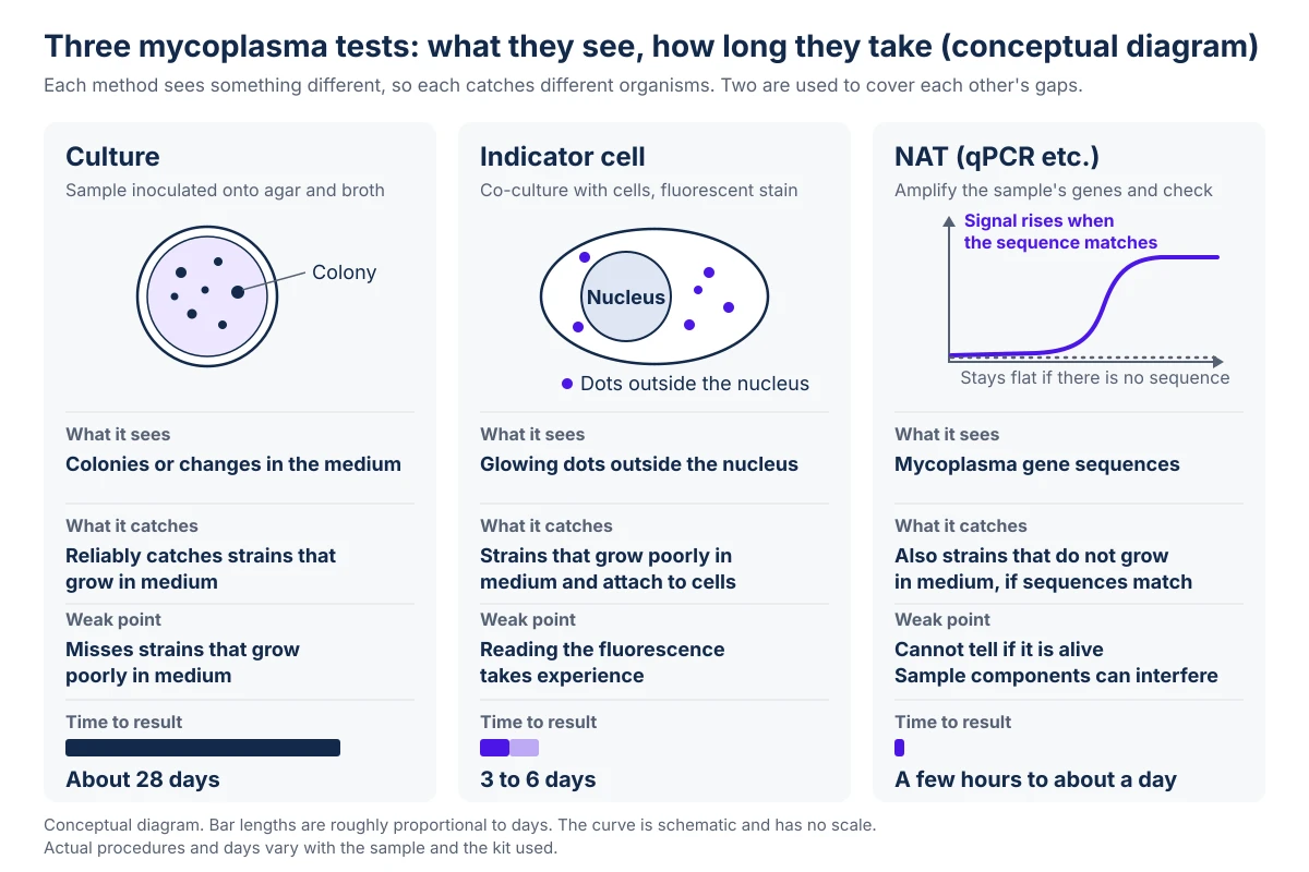
There are two classical methods because their coverage differs.
The 28 days of culture are not wasted time but the time it takes not to miss organisms that grow in medium.
What did Ph. Eur. 2.6.7 change for NAT in April 2026?
NAT can now stand on its own, if you validate it.
In the revision in force from April 1, 2026 (published in Ph. Eur. 12.2), NAT is positioned as a method that can be used by itself once validated. The detection level required of NAT is 10 CFU/mL or less, or less than 100 GC/mL. CFU is the number of colonies that grew in medium, and GC is the number of genome copies. The two do not agree for the same sample. NAT does not culture, so it gives no CFU and reports GC.
GC also counts the DNA of dead organisms. For that reason, the reference materials used to confirm the detection level now have a cap on their GC-to-CFU ratio. Running an external positive control alongside the sample is also required.
When someone says a NAT detects 10 CFU/mL, ask which reference material defined that CFU and what its GC-to-CFU ratio is.
How do you choose? Shelf life first, then sample stage
The product's shelf life decides before method performance does.
For a product such as an autologous cell therapy, where collection to administration moves in days, waiting 28 days is not an option. NAT is the realistic choice for the release decision. For a product with a long shelf life, a classical method is easier to explain (see release testing for cell therapy).
Next, split the methods by sample stage. A cell bank is a raw material used for years, and a miss contaminates every later lot. Put coverage ahead of time: combine culture and indicator cell, and add NAT if needed (see cell banks (in Japanese)). In-process and final product samples take speed, matched to the product's shelf life.
For NAT reagents, there are qPCR kits such as Thermo Fisher Scientific's MycoSEQ and Sartorius's Microsart ATMP Mycoplasma. PCR instruments come from several makers, including Roche Diagnostics' LightCycler 480 and Bio-Rad's CFX Opus. For routine monitoring of cultures there are also simple methods based on an enzyme reaction, such as Lonza's MycoAlert. Even within one maker, uses are split: Sartorius's Microsart RESEARCH Mycoplasma is for research and in-process control, while Microsart ATMP Mycoplasma is for products such as cell therapies.
Narrow the candidates by the product's shelf life, then split coverage and speed by sample stage, and you can explain why you chose as you did.
Where does a well-chosen method still fail?
In operation. Even after the method is chosen, five things break the test.
The first is using a research-use kit as it stands for release decisions. A research kit may come without validation on your product samples or documents that follow the pharmacopeial requirements.
The second is testing only the supernatant. Mycoplasma attaches to cells or gets inside them, so test the cells too.
The third is not checking for inhibition. Cell therapy samples contain many components that block the reaction, such as cells, serum components and cryoprotectants. Blocked amplification gives a false negative, so run an internal control in every run to show the reaction worked.
The fourth is not looking into the reference standard. When someone says "it detects down to 10 CFU/mL," the standard that defined that CFU changes the amount of genome the instrument is actually seeing.
The fifth is having no procedure for after a positive. A NAT positive does not distinguish live from dead. Will you follow it with culture, retest, and what happens to the lot? If a positive comes up before you have decided, the time spent deciding uses up the product's shelf life.
Release testing is the last gate, not the main defense. The controls that keep mycoplasma out come first: incoming raw material testing, operator flow and environmental control (see environmental monitoring and media fill (in Japanese)).
Fix the operating premises before the choice of method, because they are what hold up the test's reliability.
Checks you can run tomorrow
Look at your own test, or one you are about to introduce, and ask the following.
- Do you have a table that puts the product's shelf life next to the days each method needs to give a result?
- Have you assigned a method to each sample stage (cell bank, in-process, final product)?
- Does your NAT test both the cells and the supernatant, with an internal control and an external positive control in every run?
- Do you record both the GC and the CFU values of the reference material that shows the detection level?
- When a positive appears, does the procedure say who does what?
- Is the kit one that was validated on product samples, not a research-use kit?
Test kits and contract labs can be compared in the mycoplasma test directory, and PCR instruments in the real-time PCR directory. The thinking behind sterility testing is in sterility testing and rapid microbiological methods, and the differences between autologous and allogeneic manufacturing are in autologous and allogeneic cell therapy (in Japanese).
Sources
- EPC adopts mycoplasmas general chapter and monographs, updated to incorporate latest analytical developments (revised 2.6.7 published in Ph. Eur. 12.2, in force April 1, 2026). EDQM, Oct 2026.
- European Pharmacopoeia 2.6.7 Mycoplasmas (culture, indicator cell, NAT detection levels, external positive control, GC/CFU ratio of reference materials; paid text). EDQM, Apr 2026.
- Assessing the prevalence of mycoplasma contamination in cell culture via a survey of NCBI's RNA-seq archive. Nucleic Acids Research (Olarerin-George AO, Hogenesch JB; 2015;43(5):2535-2542), 2015.
- USP General Chapter <63> Mycoplasma Tests (paid text in USP-NF). United States Pharmacopeia (USP), Oct 2026.
- Japanese Pharmacopoeia: sterility test, mycoplasma test and endotoxin test (Japanese). PMDA, Oct 2026.