Japan guide · Gene therapy process guide
AAV manufacturing process: transfection to empty capsids
Photo: Eula Xandrea Dimapilis / Unsplash
One dose of Zolgensma, the gene therapy for infant spinal muscular atrophy, is 1.1×10¹⁴ vg per kilogram, given once. Here vg means vector genomes, the unit that counts virus particles carrying a gene. A 10 kg child needs about 10¹⁵ vg. Even if you assume a titer near the top of what has been reported, one 200 L bioreactor run leaves only a few patients' worth after purification.
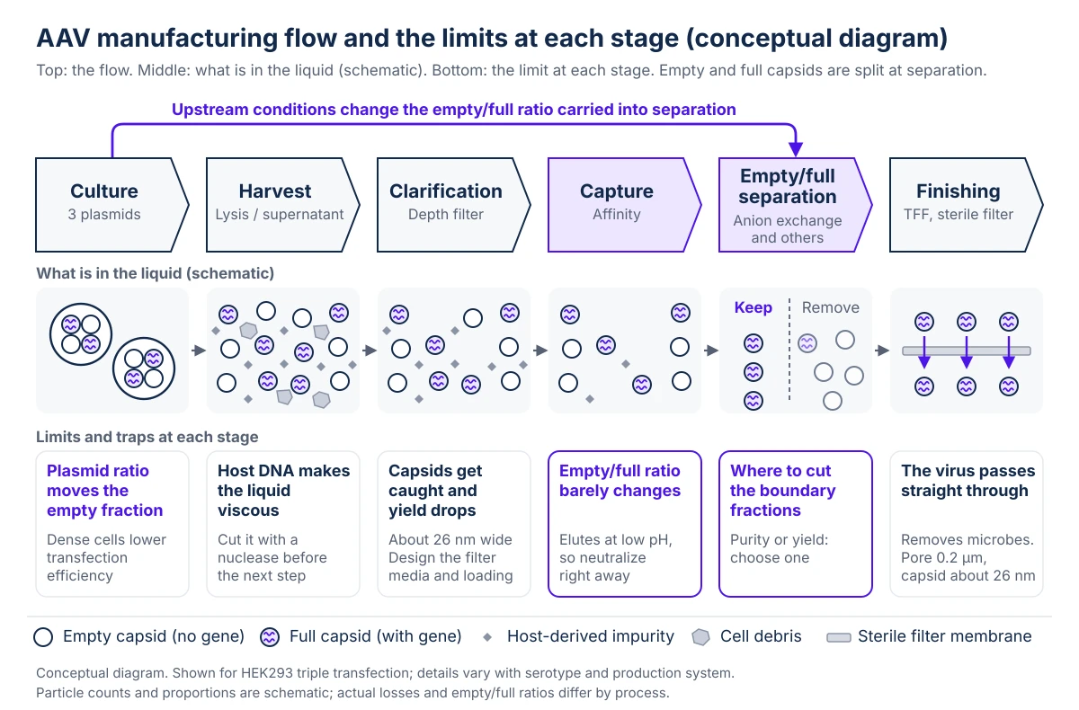
That is why AAV (adeno-associated virus) manufacturing is a yield problem before it is anything else. The sequence is fixed in outline: put three plasmids into cells so they make virus, harvest it, capture the capsids (the protein shells), and remove the empty capsids that carry no gene. Unlike antibodies, there is no standard template, because serotype and production system change the details. What each step loses becomes, directly, the number of patients you can supply.
If you buy for an AAV process, this guide shows which consumables (reagents, resins, filters) touch yield. If you work in QA, it shows where empty capsids and residuals are controlled, so a CDMO's explanation becomes easier to read.
Why does AAV volume become the bottleneck?
Systemic AAV dosing assumes very large quantities. Most approved products given systemically are dosed on the order of 10¹³ to 10¹⁴ vg per kilogram of body weight. Products injected directly into the eye or the central nervous system use a lower order of magnitude, but systemic delivery sits at this level.
In antibody manufacturing, a few percentage points of yield are a cost question. In AAV they decide how many patients one batch can treat. Every purification step erodes recovery, and empty/full separation is the costliest, because the boundary fractions are thrown away.
Approved products are not all made the same way. Zolgensma is made in HEK293 cells (human embryonic kidney cells) carrying three plasmids. Roctavian, for hemophilia A in adults, is made in Sf9 insect cells in which a baculovirus delivers the genes. The insect system needs one more step, removing the leftover recombinant baculovirus. The rest of this guide follows the most standard route, HEK293 triple transfection. For the choice between suspension and adherent culture, see AAV platform selection (in Japanese). For transient versus stable producer cell lines, see AAV production systems (in Japanese).
A few points of yield that an antibody process can absorb become, in AAV, a count of patients.
Plasmid ratios set upstream decide how hard separation is downstream
Culture starts by splitting the genes across three plasmids. The first carries the transgene, flanked by ITRs (inverted terminal repeats, sequences the virus needs for replication). The second carries cap, which builds the capsid, and rep, which packs the genome into it. The third carries helper functions derived from adenovirus, which support replication. One virus originally held all of these functions, and splitting them lowers the chance that a wild-type virus re-forms (see triple transfection (in Japanese)).
Transfection reagents include PEIpro (Polyplus / Sartorius) and AAV-MAX (Thermo Fisher Scientific). A denser culture does not always give more total product. Transfection is usually done at a fairly low cell density, because efficiency per cell falls as density rises.
The ratio among the three plasmids also matters. Changing it shifts the balance between how much capsid is made and how well genomes are packed, and that moves the fraction of empty capsids. All three plasmids must be GMP grade (made to pharmaceutical manufacturing standards), and their delivery date often drives the schedule. The CDMO selection guide (in Japanese) covers what to ask.
Set the plasmid ratio with the empty-capsid load in mind, because it fixes how much separation work you face later.
Affinity capture is efficient, but it cannot tell empty capsids from full ones
How you harvest depends on the serotype. A serotype that tends to stay inside the cell, such as AAV2, is collected as cells and lysed with detergent to release the contents. Serotypes that tend to leave the cell, such as AAV8 and AAV9, can be processed from the culture supernatant as it is. Either way, host DNA from broken cells makes the liquid viscous, so a nuclease such as Benzonase cuts it before the next step.
Clarification protects the columns that follow. It relies mainly on depth filters (thick filter media) to remove cells and debris. If the capsids, about 26 nm across, are caught along with the debris, recovery drops. The choice of filter media and the loading design show up directly in yield.
The first chromatography step is affinity capture. The resin grabs the capsid surface. A resin such as POROS CaptureSelect AAVX (Thermo Fisher Scientific) binds several serotypes, so one resin can serve different products (see AAV affinity resins). Its place in the process is close to that of Protein A in antibody purification. One step removes most host-derived impurities, which makes it an efficient one. Elution is at low pH, and capsids are unstable in acid, so fix the neutralization procedure right after elution, including how long it may take.
The step has a limit. The resin grabs the capsid surface and does not see whether anything is inside.
Capture leaves the empty-to-full ratio almost unchanged, so plan a dedicated separation step.
Empty/full separation is a cut-point decision, and the starting ratio comes from upstream
The real separation comes after capture. Empty and full capsids have the same shell protein and the same diameter. Only the contents differ. Empty capsids cannot deliver a gene, yet they add to the amount of particles dosed. You are sorting particles that look identical from outside by a small difference in the genome inside.
There are two approaches. Ultracentrifugation uses the density difference. It resolves well but does not suit large scale. Anion exchange uses the slight difference in surface charge that the genome creates. It runs on ordinary equipment and resin, fits manufacturing, and is the standard choice (see anion exchange for full/empty separation (in Japanese)). With either method you still have to decide where to cut the boundary fractions. Cut for purity and yield falls. Cut for yield and empty capsids remain.
The empty/full ratio is not set by the separation step alone. Plasmid ratio, cell density and harvest timing all move the ratio at the end of capture. If the ratio entering separation is good, the same cut reaches higher purity. Treating empty-capsid control as a downstream job leaves upstream gains unused.
The reference method for measuring the ratio is analytical ultracentrifugation (AUC; see the AUC product guide). One run is reported to take about 6 hours and needs a lot of sample. For in-process control, some programs pair it with faster methods such as mass photometry, which measures in minutes. Different methods measure different things, so read a specification number together with its method.
Quote an empty/full number only together with the method that produced it.
The sterile filter at the end removes microbes, not the virus
The last stage adjusts concentration and composition. A TFF system concentrates the product and exchanges it into the formulation buffer. Then the product goes through a sterile filter, and here is a trap. The filter pore is 0.2 µm and the capsid is about 26 nm, so the virus passes straight through.
This step removes microorganisms. It does not remove the virus.
Make sure everyone on the team knows that the sterile filter takes out microorganisms and lets the virus pass.
Titers compare only against a shared reference standard
Quality testing is built on several axes: genome quantity, capsid quantity, the empty/full ratio, residual DNA, residual plasmid, residual nuclease, host cell protein, and the titer for infectivity or expression. Titer numbers do not line up when each site measures differently. The industry therefore uses reference materials supplied by ATCC, AAV2 (VR-1616) and AAV8 (VR-1816), as a common yardstick.
Even so, when the AAV8 material was measured at three sites, the variation in infectious titer was larger than the mean. When you compare a CDMO's titer with your own test, measure the same standard on both sides and check.
Test data are the basis of the process itself. In August 2019, FDA announced that some animal-study data in Zolgensma's marketing application had been manipulated. The developer, AveXis, had told the agency in June. FDA said the problem was confined to part of the data supporting process development, and it let the product stay on the market. It did fault the company for knowing before approval and disclosing only afterward. Reliable analysis and records are questioned after approval as well.
For process-derived impurities and safety, see AAV impurities and liver toxicity (in Japanese). For the whole testing picture, see gene therapy quality control (in Japanese).
Run the same reference standard on both sides before you compare a CDMO's titer with your own.
Seven things to confirm with the developer or CDMO
Work through these in order for any AAV project.
- Confirm the serotype and the production system (HEK293 triple transfection or insect cells). Harvest method and resin choice both depend on it.
- Ask when the three plasmids and the transfection reagent will be delivered, and whether they are GMP grade.
- Ask whether the capture resin has a track record with your serotype, and whether the neutralization step after elution has a time limit.
- Check in the specification which method separates empty from full capsids, what ratio is the limit, and how that ratio is measured.
- Get the recovery for every step and the total yield. Knowing where you lose the most tells you where to improve.
- Share inside your company what the sterile filter does and does not remove.
- Agree with the CDMO on a common titer reference standard.
For purchasing or sales, a good opening question is "Which serotypes have you made, by which method, and up to what scale?" It makes the conversation concrete at once. Resins for the separation step can be compared in the product directory under anion exchange resins and membrane chromatography. The whole process on one page is the AAV process map.
Sources
- Chemistry, Manufacturing, and Control (CMC) Information for Human Gene Therapy Investigational New Drug Applications (INDs). FDA, Jan 2020.
- Zolgensma EPAR Product Information. European Medicines Agency, Oct 2026.
- Bioprocessing and Scale-Up of Suspension HEK293 Cells for AAV Manufacturing. Merck, Oct 2026.
- Evaluation of recombinant baculovirus clearance during rAAV production in Sf9 cells using a newly developed fluorescent-TCID50 assay. Frontiers in Medicine, 2024.
- Zolgensma: We all know the price, but how is it made?. BioProcess International, Oct 2026.
- Quantification of Empty, Partially Filled and Full Adeno-Associated Virus Vectors Using Mass Photometry. International Journal of Molecular Sciences, Jan 2024.
- Stability of the adeno-associated virus 8 reference standard material. INSERM (HAL open archive), Oct 2026.
- FDA assessing data manipulation concerns with gene therapy product. Pharmacy Times, Aug 2019.